Diagram Of Backbone - Biology - All About Bats / Human skeleton with each bone name. Cervical spine is the spine in the neck area. The spinal cord begins at the bottom of the brain stem (at the area called the medulla oblongata) and ends in the lower back, as it tapers to form a cone called the conus medullaris. All the images are in vector format, allowing an optimal web display with zoom and shifting of the anatomical images. The 12 vertebral bodies in the upper back make up the thoracic spine. The bones of the pelvis and lower back work together to support the body's weight, anchor the abdominal and hip muscles, and protect the delicate vital organs of the vertebral and abdominopelvic cavities.
The vertebral column, also known as the backbone or spine, is part of the axial skeleton.the vertebral column is the defining characteristic of a vertebrate in which the notochord (a flexible rod of uniform composition) found in all chordates has been replaced by a segmented series of bone: The spinal cord is an extension of the central nervous system (cns), which consists of the brain and spinal cord. The thoracic spine is basically a strong cage and it is designed to protect the vital organs of the heart and lungs. The spinal column (vertebral column or backbone) provides both structural and nervous system support for your entire body. Backbone diagram with vertebrae, disks and nerves.
Mengenal Jaringan Backbone dan Manfaatnya - Telko.id from telko.id Long bone diagram labeled colored. The vertebrae are divided into five sections: Human back muscles and bones. We think this is the most useful anatomy picture that you need. The spinal cord is an extension of the central nervous system (cns), which consists of the brain and spinal cord. The spinal cord begins at the bottom of the brain stem (at the area called the medulla oblongata) and ends in the lower back, as it tapers to form a cone called the conus medullaris. As you can see in the vertebrae diagram above, the human spine consists of 33 vertebrae in total; The thoracic spine helps keep the body upright and stable.
Anatomical diagrams of the spine and back.
Spine diagrams the human spine consists of 33 vertebrae: The back is the body region between the neck and the gluteal regions. Vertebrae separated by intervertebral discs. Simply line up the vertebral level with the possible symptoms and you will see some surprising connections of symptoms that relate to your spine. This section is made up of 7 vertebrae (abbr. The intervertebral foramen (neural passageways. Long bone diagram labeled colored 12 photos of the long bone diagram labeled colored , bone. The vertebral column, also known as the backbone or spine, is part of the axial skeleton.the vertebral column is the defining characteristic of a vertebrate in which the notochord (a flexible rod of uniform composition) found in all chordates has been replaced by a segmented series of bone: The 12 vertebral bodies in the upper back make up the thoracic spine. The trapezius or trapezoid muscles are two paired muscles that extend from the base of the thoracic vertebrae in the spine to the occipital bone and run out to the spine of the scapula. Network diagram software backbone network backbone network diagraming. The pedicles are longer and wider than those in the thoracic spine. Backbone diagram with vertebrae, disks and nerves.
Spine diagrams the human spine consists of 33 vertebrae: This section is made up of 12 vertebrae (abbr. The spine anatomy is a complex structure. Vertebrae separated by intervertebral discs. It comprises the vertebral column (spine) and two compartments of back muscles;
Back Bones Diagram / Vintage Anatomy Skeleton Images - The ... from www.101diagrams.com Network diagram software backbone network backbone network diagraming. The spine diagram shown below, consists of many bones or vertebrae,soft discs,the spinal cord, and spinal nerves. Vertebrae separated by intervertebral discs. The muscles of the lower back help stabilize, rotate, flex, and extend the spinal column, which is a bony tower of 24 vertebrae that gives the body structure and houses the spinal cord. C1 to c7) and are the neck bones of the spinal column. The vertebrae are divided into five sections: Human back muscles and bones 12 photos of the human back muscles and bones human back muscles and bones, bone, human back muscles and bones. Spine diagrams the human spine consists of 33 vertebrae:
The trapezius or trapezoid muscles are two paired muscles that extend from the base of the thoracic vertebrae in the spine to the occipital bone and run out to the spine of the scapula.
Anatomy of human body seen from behind. It comprises the vertebral column (spine) and two compartments of back muscles; 24 are considered to be part of the upper spine, whilst the other 11 are found in the sacrum & coccyx. We hope this picture anatomy of back muscles diagram can help you study and research. The vertebral column of the lower back includes the five lumbar vertebrae, the sacrum, and the coccyx. Human skeleton with each bone name The spine diagram shown below, consists of many bones or vertebrae,soft discs,the spinal cord, and spinal nerves. Lower back vertebrae (5) (lumbar vertebrae) back of skull (occipital bone) fused vertebrae (5) (sacrum) hand bones (metacarpals) finger bones (phalanges) heel bone (calcaneus) skull (cranium) backbone. We think this is the most useful anatomy picture that you need. The spinal column (vertebral column or backbone) provides both structural and nervous system support for your entire body. Anatomically, the spinal cord runs from the top of the highest neck bone (the c1 vertebra) to. The vertebral column houses the spinal canal, a cavity that. Spine diagrams the human spine consists of 33 vertebrae:
The spinous processes are horizontal and more squared in shape. The thoracic spine is the middle part of the spine, connecting the cervical and lumbar spine. Backbone diagram with vertebrae, disks and nerves. Cervical spine is the spine in the neck area. Vertebrae separated by intervertebral discs.
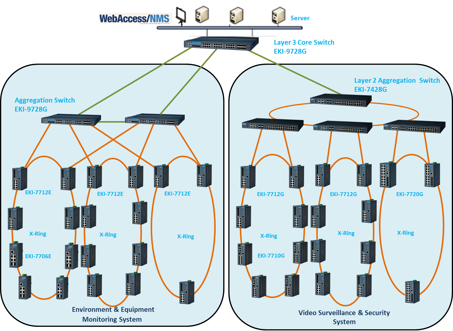
Building an Effective Ethernet Backbone Network for China ... from advcloudfiles.advantech.com The vertebral column, also known as the backbone or spine, is part of the axial skeleton.the vertebral column is the defining characteristic of a vertebrate in which the notochord (a flexible rod of uniform composition) found in all chordates has been replaced by a segmented series of bone: The back is the body region between the neck and the gluteal regions. Human bone anatomy 3d 9 photos of the human bone anatomy 3d human bone anatomy diagram, human bone anatomy game, human bone anatomy pdf, human bone anatomy quiz, human leg bone anatomy, maxillary bone anatomy 3d, muscle and bone anatomy 3d, temporal bone anatomy 3d, human anatomy, human bone anatomy diagram, human bone anatomy game, … On the chart below you will see 4 columns (vertebral level, nerve root, innervation, and possible symptoms). We think this is the most useful anatomy picture that you need. T1 to t12) and are the chest back bones of the spinal column. The vertebrae, which stack like spools of thread, support the back and protect the spinal cord. Full woman patient body from back.
The muscles of the lower back help stabilize, rotate, flex, and extend the spinal column, which is a bony tower of 24 vertebrae that gives the body structure and houses the spinal cord.
Anatomical diagrams of the spine and back. On the chart below you will see 4 columns (vertebral level, nerve root, innervation, and possible symptoms). Anatomy of human body seen from behind. Long bone diagram labeled colored. T1 to t12) and are the chest back bones of the spinal column. For more anatomy content please follow us and visit our website: The spinal cord begins at the bottom of the brain stem (at the area called the medulla oblongata) and ends in the lower back, as it tapers to form a cone called the conus medullaris. These vertebrae bear much of the body's weight and related biomechanical stress. All the images are in vector format, allowing an optimal web display with zoom and shifting of the anatomical images. The normal curvature of the cervical vertebrae is 'inwards' (lordosis). Anatomynote.com found anatomy of back muscles diagram from plenty of anatomical pictures on the internet. Cervical spine is the spine in the neck area. Lower back vertebrae (5) (lumbar vertebrae) back of skull (occipital bone) fused vertebrae (5) (sacrum) hand bones (metacarpals) finger bones (phalanges) heel bone (calcaneus) skull (cranium) backbone.